Further Breast Assessment: The Role of Mammography, Ultrasound and MRI
When a breast screening mammogram or clinical examination identifies an area that requires further investigation, additional imaging may be recommended to obtain more information.
Mammography, ultrasound and magnetic resonance imaging (MRI) provide different views of breast tissue. Each technique has its own strengths and uses, and the choice depends on the finding being assessed and the clinical question that needs to be answered.
Understanding how these imaging methods differ can help explain why a radiologist may recommend one examination or a combination of investigations.
1. Why Might Additional Breast Imaging Be Needed?
A screening mammogram provides an initial assessment of breast tissue. However, some findings may not be fully characterised from the standard images.
Additional imaging may help the radiologist:
Examine a particular area more closely.
Determine whether a finding represents overlapping tissue or a distinct abnormality.
Assess whether a lump is fluid-filled or solid.
Evaluate the characteristics and extent of an abnormality.
Identify the most appropriate method for image-guided tissue sampling, if required.
The choice of imaging depends on the initial findings, breast characteristics, symptoms and relevant clinical history.
2. Diagnostic Mammography: Examining a Finding More Closely
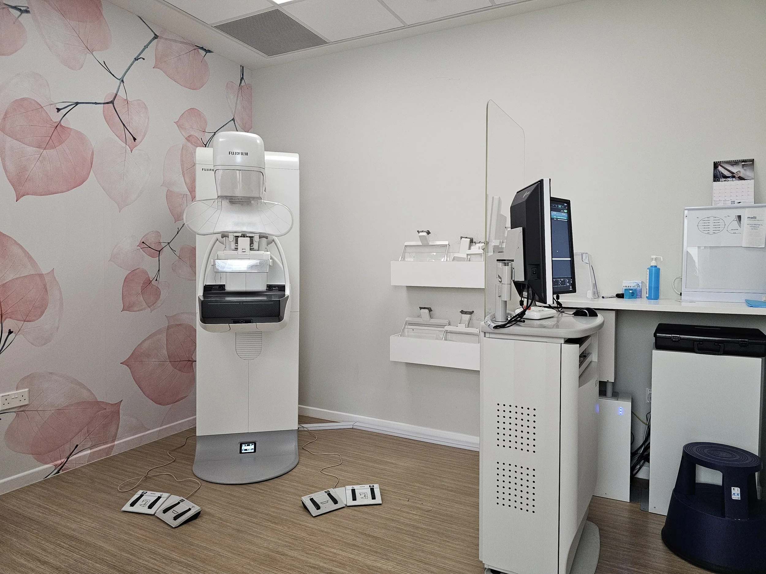
Mammography uses low-dose X-rays to produce images of breast tissue. While screening mammography is performed to look for abnormalities in women without breast symptoms, diagnostic mammography is used to investigate a specific finding or clinical concern.
Additional mammography views may include:
Spot compression views: Apply focused compression over a particular area to help separate overlapping breast tissue.
Magnification views: Provide a closer examination of small findings, particularly calcifications.
Additional angled views: Show an area from a different direction to help clarify its appearance.
These images allow the radiologist to examine the shape, margins and distribution of a finding in greater detail.
What Is 3D Mammography?
Three-dimensional mammography, also known as digital breast tomosynthesis (DBT), captures multiple low-dose X-ray images from different angles as the X-ray tube moves in an arc around the breast.
The images are reconstructed into thin sections, allowing the radiologist to examine breast tissue layer by layer. This can help reduce the effect of overlapping tissue and improve the visibility of certain abnormalities.
3D mammography can be used in breast screening and diagnostic assessment. However, it does not detect every breast abnormality and may still need to be supplemented by other imaging examinations.
Learn more about 3D Mammography at Luma
3. Breast Ultrasound: Assessing the Structure of an Abnormality
Breast ultrasound uses high-frequency sound waves to produce images of breast tissue. It does not use ionising radiation.
It is commonly used to investigate a breast lump or examine an area identified on mammography.
Ultrasound can help the radiologist assess:
Whether a lump is fluid-filled or solid.
The shape and margins of a lesion.
The internal characteristics of an abnormality.
The relationship of a finding to surrounding breast tissue.
How Does Ultrasound Complement Mammography?
Mammography and ultrasound provide different information.
For example, a mammogram may identify an area of concern, while ultrasound can help determine whether the finding corresponds to a cyst or solid lesion.
Ultrasound is also useful for guiding a biopsy needle towards an abnormality that can be clearly seen during the examination.
However, ultrasound does not replace mammography in every situation. The two methods are often complementary, and the need for each depends on the clinical findings.
Learn more about Breast Ultrasound at Luma
4. Breast MRI: Providing Additional Information in Selected Cases
Breast magnetic resonance imaging (MRI) uses a strong magnetic field, radio waves and a computer to produce detailed images of breast tissue. It does not use ionising radiation.
Most breast MRI examinations performed for cancer detection or assessment involve an intravenous contrast agent. This helps highlight areas of tissue with increased contrast enhancement.
Breast MRI may be considered in selected situations, including:
Assessing the extent of a known breast cancer: MRI may help evaluate the extent of disease within the breast and identify additional areas of concern.
Investigating selected imaging findings: It may be considered when mammography and ultrasound do not provide sufficient information to answer a particular clinical question.
Screening women at increased risk: MRI may be recommended for women with certain hereditary breast cancer gene mutations or a sufficiently high estimated lifetime risk.
Assessing specific clinical concerns: MRI may provide additional information in selected cases involving breast implants or other complex findings.
MRI is not required for every woman and does not routinely replace mammography or ultrasound. The decision depends on the clinical question and whether MRI is likely to provide useful additional information.
Learn more about Breast MRI at Luma
5. Why Might More Than One Imaging Method be Used?
Different imaging methods can provide complementary information about the same area of breast tissue.
For example:
Mammography may identify a group of calcifications that requires closer examination.
Ultrasound may help determine whether a lump is fluid-filled or solid.
MRI may provide additional information about the extent of a known cancer or a selected finding that remains unclear after other imaging.
Using more than one imaging method does not automatically mean that a finding is cancerous. It may simply be necessary to answer different aspects of the clinical question.
6. How Does the Radiologist Decide Which Imaging Examination Is Needed?
The radiologist considers the appearance of the finding and the information already available before recommending further imaging.
Factors may include:
The type, location and characteristics of the abnormality.
Whether the finding was identified during screening or diagnostic assessment.
Breast density and tissue characteristics.
Symptoms and clinical examination findings.
Previous mammograms, ultrasound or MRI examinations.
Personal and family history, where relevant.
Previous imaging is particularly useful because it allows the radiologist to determine whether a finding is new, unchanged or has developed over time.
If imaging findings remain uncertain, an image-guided biopsy may be recommended to obtain tissue for pathological examination.
Frequently Asked Questions
-
Neither examination is universally better. Mammography and ultrasound provide different information and may be used together when clinically indicated. The appropriate examination depends on the finding and the clinical question.
-
No. 3D mammography provides reconstructed sections of breast tissue, while ultrasound uses sound waves to examine the breast. They have different roles and may complement one another during diagnostic assessment.
-
MRI can provide additional information in selected situations, but it is not a replacement for mammography in every patient. The most appropriate examination depends on the clinical circumstances.
-
No. MRI uses a magnetic field and radio waves rather than ionising radiation. Most breast MRI examinations for cancer assessment use an intravenous contrast agent.
-
No. Most women do not need every type of breast imaging. The breast specialist recommends examinations based on the finding being investigated and the information needed for assessment.
Breast Imaging Services at Luma
Luma Women's Imaging Centre provides 3D mammography, breast ultrasound and breast MRI for breast screening and diagnostic assessment. The appropriate examination is selected according to the clinical question and individual circumstances.