Breast MRI Scan
Breast MRI (magnetic resonance imaging) uses a strong magnetic field, radio waves and a computer to produce detailed images of breast tissue. For women at increased risk of breast cancer, breast MRI may be recommended alongside mammography as a screening tool. It may also be used to assess the extent of a known breast cancer or investigate selected findings that require further evaluation.
At Luma Women’s Imaging Centre, breast MRI forms part of a wider range of breast imaging and diagnostic services. The choice of imaging depends on the clinical question, medical history, individual risk and findings from previous examinations.
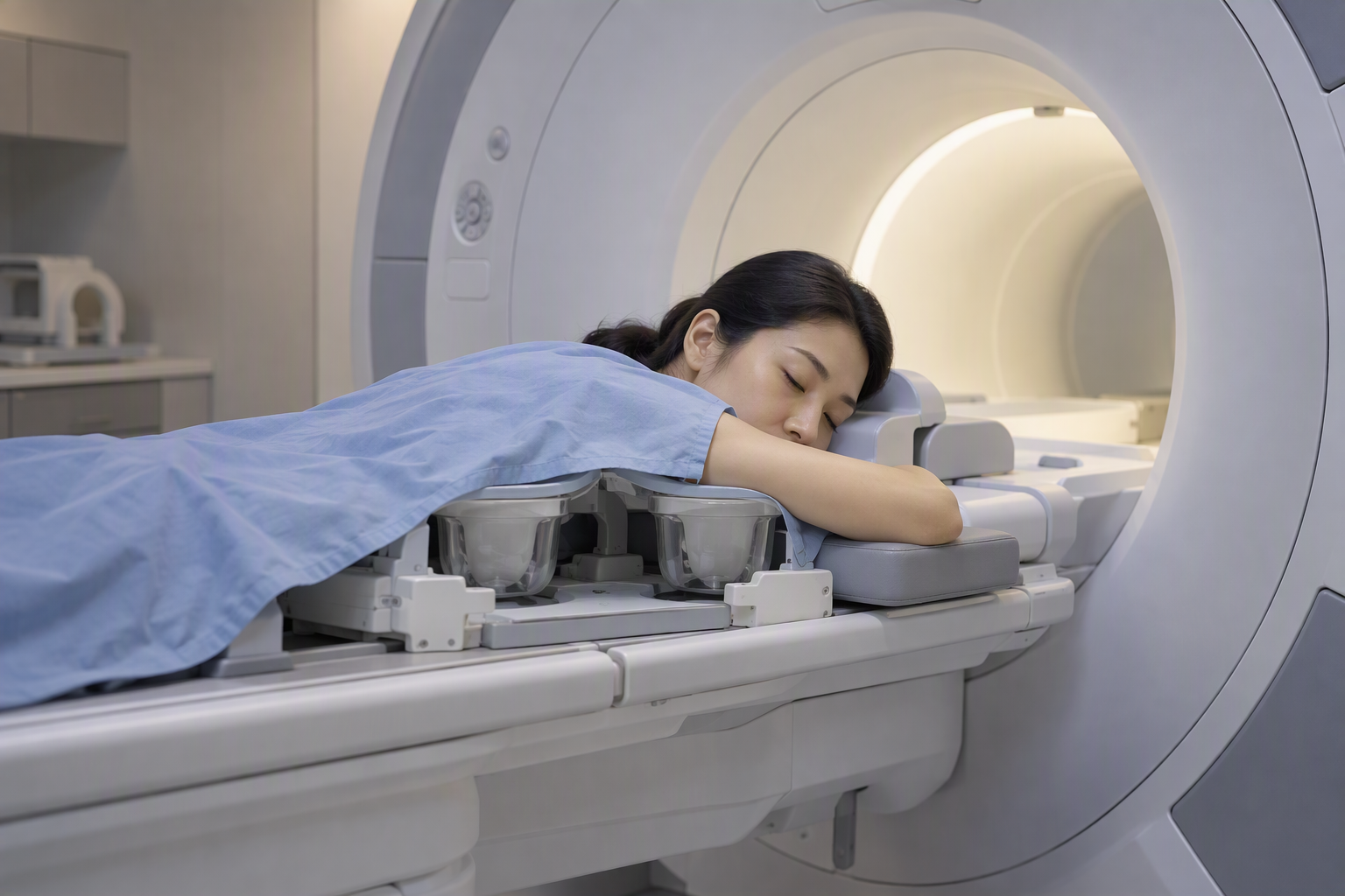
What is a Breast MRI Scan?
Unlike mammography, which uses low-dose X-rays, MRI does not use ionising radiation. It provides detailed images of soft tissue and can reveal some abnormalities that may not be clearly seen on mammography or ultrasound.
Most breast MRI examinations performed to detect or assess breast cancer use an intravenous contrast agent to help highlight areas of abnormal tissue. The need for contrast depends on the purpose of the examination.
Breast MRI complements rather than routinely replaces mammography or ultrasound. It is not necessary for every woman and should be recommended according to individual clinical needs.
Breast MRI for Breast Cancer Screening
For women at increased risk of breast cancer, breast MRI may be recommended alongside mammography to improve cancer detection. MRI can identify some breast cancers that may not be visible on mammography, although it can also identify findings that require additional investigation but are not cancer.
Breast MRI screening may be considered for:
Women with hereditary breast cancer gene mutations: Certain mutations, including BRCA1 and BRCA2, are associated with an increased risk of breast cancer and may warrant enhanced screening.
Women with a significant family history of breast cancer: MRI may be recommended when an assessment of personal and family history indicates a sufficiently high lifetime risk.
Women with very dense breast tissue: Dense tissue can make abnormalities more difficult to detect on mammography. Supplemental MRI may be considered depending on breast density, overall risk and applicable clinical guidance.
The decision to undergo breast MRI for screening should be made in consultation with a doctor, taking into account individual risk factors, previous imaging findings and the potential benefits and limitations of additional screening.
Breast MRI does not routinely replace mammography. The recommended screening approach depends on each woman's clinical circumstances.
When Is a Breast MRI Recommended?
Beyond screening for women at increased risk, a doctor or radiologist may recommend breast MRI in selected circumstances, including:
Assessing a known breast cancer: MRI may help evaluate the extent of a newly diagnosed cancer and identify additional areas that may require further assessment.
Investigating selected imaging findings: MRI may be considered when mammography or ultrasound does not provide enough information to resolve a specific clinical question.
Monitoring response to treatment: In some cases, MRI helps assess how a breast cancer has responded to treatment before surgery.
The suitability of MRI depends on the individual clinical situation. Having dense breast tissue or breast implants does not automatically mean that MRI is required for breast cancer screening.
What Happens During Your Breast MRI Scan?
A breast MRI scan is generally painless, although lying still in the scanner may feel uncomfortable for some people.
Getting into position: You will change into suitable clothing and lie face down on a specially designed examination table, with your breasts positioned within an opening in the table.
During the scan: The table moves into the MRI scanner. You will need to remain as still as possible to help produce clear images. The machine makes repetitive sounds, and earplugs or headphones may be provided.
Contrast injection: For most breast MRI examinations used to detect or assess breast cancer, a contrast agent is injected into a vein in your arm during the scan.
Communication and comfort: The radiographer monitors the examination from a nearby room and can communicate with you through an intercom. You can alert the team if you experience discomfort.
The scan itself typically takes around 30–45 minutes, although the total appointment may take longer depending on the examination and any additional imaging required.
What Happens After the Scan?
The images will be reviewed by a radiologist, who will prepare a report for the referring doctor or healthcare professional according to the centre's reporting workflow.
Depending on the findings, you may be advised that no further investigation is needed, that follow-up imaging should be considered, or that additional assessment or a biopsy may be appropriate.
An area that appears abnormal on MRI does not necessarily indicate cancer. Further tests may sometimes be needed to clarify the finding.
Frequently Asked Questions About Breast MRI
-
No. Breast MRI and mammography provide complementary information. For women at higher risk of breast cancer, MRI may be recommended in addition to mammography. The appropriate combination depends on individual risk and clinical circumstances.
-
MRI does not use ionising radiation. However, its strong magnetic field means that certain implanted devices or metal objects require careful safety assessment. If contrast is needed, the team will also consider relevant medical history, including kidney problems and previous contrast reactions.
-
No. Contrast is used in most breast MRI examinations performed to detect or assess breast cancer. However, MRI performed specifically to evaluate silicone breast implant integrity generally does not require intravenous contrast. The protocol depends on the clinical purpose of the scan.
-
Yes. Breast MRI can be used to assess the integrity of silicone breast implants and detect signs of rupture, including ruptures that may not cause noticeable symptoms.
The appropriate imaging method and timing depend on the type of implant, how long it has been in place and individual circumstances. If you have breast implants and are concerned about a possible complication, consult your doctor about the appropriate examination.
-
An abnormal finding does not necessarily mean cancer. Depending on the appearance and clinical context, the radiologist may recommend additional imaging, follow-up or a biopsy to establish the nature of the finding.
Breast MRI Services at Luma
Luma provides breast imaging and diagnostic services to support further investigation when clinically indicated. Our radiology team works with referring healthcare professionals to help ensure that imaging findings are appropriately evaluated.
For referring doctors: For examination requests or referral enquiries, please use our Radiology Request Form or contact our team.
For patients: If your doctor has recommended a breast MRI or you would like to enquire about the examination, contact Luma for further information.
Learn more about breast MRI
For a convenient overview of breast MRI, including what the examination involves, download our brochure.
For detailed instructions on preparing for your examination, please refer to our Breast MRI Preparation Guide.