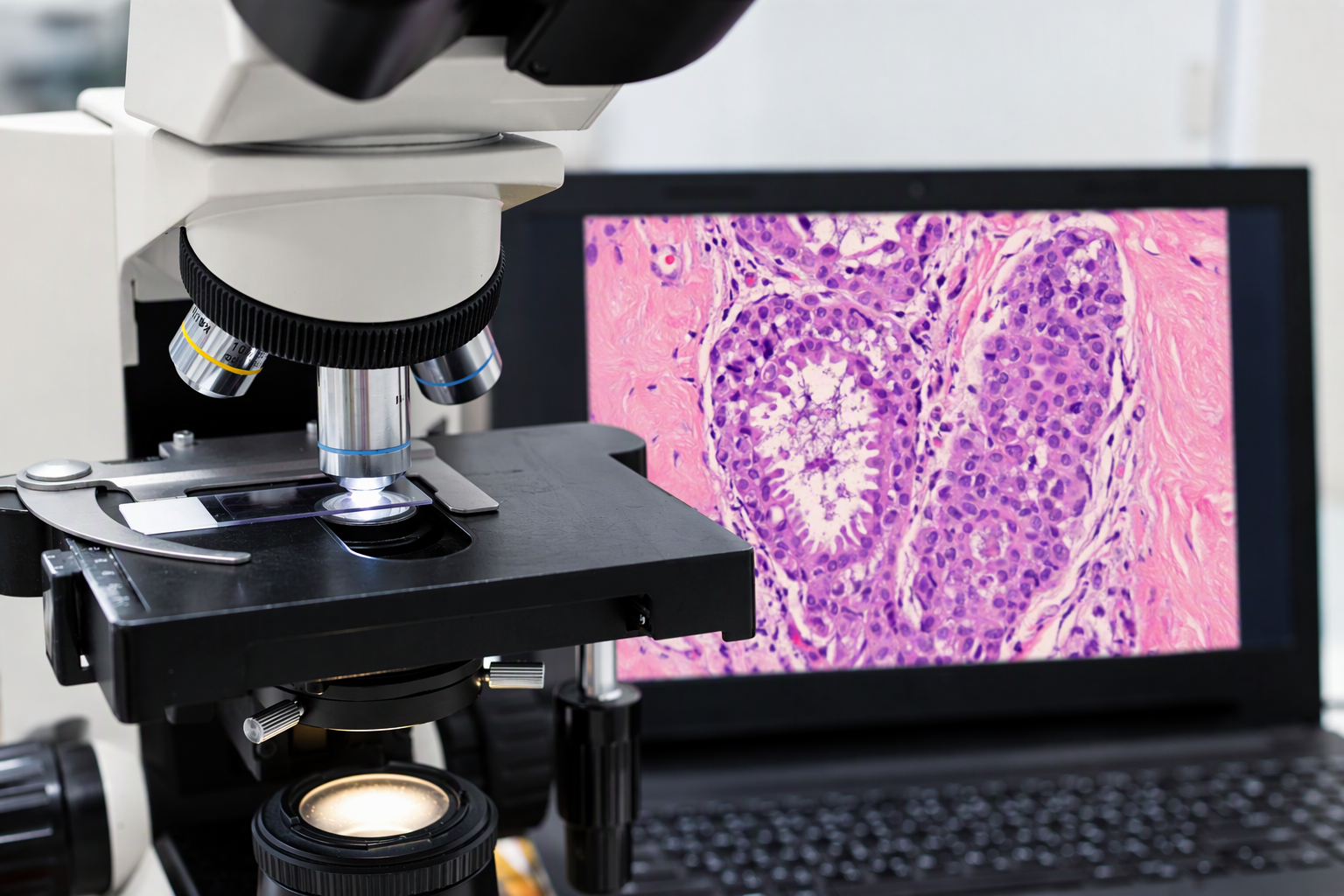
Image-Guided Breast Biopsy: From Tissue Sampling to Pathology Assessment
When a breast abnormality requires tissue examination, an image-guided biopsy may be recommended. This procedure uses imaging to guide a needle towards the area of concern so that tissue or cell samples can be collected for pathological examination.
The imaging technique used depends on how the abnormality is seen and where it is located. After the biopsy, a pathologist examines the collected samples to determine the nature of the finding.
Understanding the relationship between imaging, biopsy and pathology helps explain how these different parts of the diagnostic process contribute to establishing a diagnosis.
1. When is an Image-Guided Breast Biopsy Recommended?
A breast biopsy may be recommended when an abnormality identified through imaging or clinical examination requires tissue examination.
This may include:
A breast mass with imaging characteristics that require further investigation.
An area of architectural distortion on a mammogram.
Calcifications that need pathological assessment.
An abnormality identified on ultrasound or MRI.
A finding that cannot be confidently characterised through imaging alone.
Not every breast abnormality requires a biopsy. Some findings have characteristic benign appearances and may only require imaging follow-up.
When tissue examination is needed, the breast specialist considers the appearance and location of the abnormality to determine the most suitable biopsy technique.
2. What Are the Different Types of Breast Biopsy?
Several techniques can be used to obtain breast tissue or cell samples. The choice depends on the nature of the abnormality and the information required for diagnosis.
Core Needle Biopsy (CNB)
A core needle biopsy uses a hollow needle to remove small samples of breast tissue for examination.
Local anaesthesia is usually administered to numb the area before the procedure. The needle is guided towards the abnormality using ultrasound, mammography or MRI, depending on how the finding is visualised.
The samples preserve the structure of the tissue, allowing the pathologist to examine the cells and their arrangement.
Vacuum-Assisted Biopsy (VAB)
A vacuum-assisted biopsy uses a specialised needle connected to a vacuum device to collect multiple tissue samples through a small skin incision.
The technique allows several samples to be obtained through a single needle insertion. It may be used for selected breast abnormalities, including certain small lesions and areas of calcification.
Depending on the size and nature of the abnormality, VAB may also be used to remove certain small, benign breast lesions. It is primarily a diagnostic procedure and is not a substitute for surgery to remove breast cancer.
Fine Needle Aspiration (FNA)
Fine needle aspiration uses a thin needle to collect cells or fluid from a breast lump or other targeted area.
It may be suitable for selected cases, including the assessment of certain cysts or lumps. Unlike a core needle biopsy, which collects small pieces of tissue, FNA provides individual cells or fluid for examination.
The choice between FNA and a core needle biopsy depends on the clinical question and the information needed.
3. How is the Biopsy Guided by Imaging?
Most breast needle biopsies are performed using imaging guidance. This allows the radiologist to visualise the abnormality and guide the needle towards the intended area.
The guidance technique is selected according to which imaging examination best demonstrates the finding.
Ultrasound-Guided Biopsy
When an abnormality can be clearly seen on ultrasound, the radiologist uses real-time ultrasound images to guide the needle into the targeted area.
This technique is commonly used for breast lumps and other abnormalities that are visible on ultrasound.
Mammography-Guided Biopsy
Some findings, particularly certain areas of microcalcification, may be visible on mammography but not clearly demonstrated on ultrasound.
In these cases, stereotactic or mammography-guided biopsy may be recommended. The imaging system helps the radiologist determine the location of the abnormality and guide the biopsy needle.
MRI-Guided Biopsy
Some breast abnormalities are visible on MRI but cannot be adequately targeted using ultrasound or mammography.
When tissue sampling is required, an MRI-guided biopsy may be considered. The procedure is performed using MRI images to guide the needle towards the area of concern.
MRI-guided biopsy requires specialised equipment and expertise. It provides an option for sampling selected abnormalities that are best demonstrated on MRI.
4. What Happens During an Image-Guided Biopsy?
Although the details vary according to the biopsy technique, the procedure generally involves several steps:
Positioning: You will be positioned according to the imaging technique being used.
Preparation: The skin over the biopsy site is cleaned, and local anaesthesia is administered where appropriate.
Image guidance: The radiologist uses ultrasound, mammography or MRI to locate the abnormality and guide the needle.
Tissue sampling: The required tissue or cell samples are collected.
Aftercare: Pressure is applied to the biopsy site to reduce bleeding, followed by a dressing.
Some biopsy procedures may be performed with sedation when clinically appropriate. The recommended approach, preparation and recovery arrangements will be explained before the procedure.
Learn more about Breast Biopsy at Luma
5. What Happens to the Tissue Samples After a Biopsy?
Once collected, the tissue samples are sent for pathological examination.
A pathologist examines the samples under a microscope to assess the structure and characteristics of the cells and tissue.
The pathology report may describe:
The type of breast tissue or lesion identified.
Whether the findings are benign, atypical or malignant.
The specific characteristics of any abnormal cells.
Additional tests or biomarkers, where indicated.
For certain breast cancer diagnoses, further testing may be performed to assess biomarkers that help characterise the tumour and inform treatment decisions.
Breast Cancer Biomarker Assessment
Commonly assessed breast cancer biomarkers include:
Oestrogen receptor (ER): Determines whether the cancer cells express receptors for oestrogen.
Progesterone receptor (PR): Determines whether the cancer cells express receptors for progesterone.
HER2: Assesses HER2 protein expression and, where indicated, gene amplification.
Ki-67: Provides an indication of the proportion of tumour cells undergoing proliferation.
These results are interpreted alongside the histological findings and other clinical information. They help doctors understand the characteristics of a breast cancer and consider appropriate treatment options.
6. Why Must Imaging and Pathology Findings Be Considered Together?
Imaging and pathology provide different but complementary information.
Radiologists assess the appearance, location and characteristics of an abnormality through mammography, ultrasound or MRI. Pathologists examine the collected tissue or cells to determine the nature of the finding.
The two sets of findings need to be considered together to establish whether the pathology result adequately explains the abnormality seen on imaging. This process is known as radiology-pathology correlation.
What Is Radiology-Pathology Concordance?
When the pathology findings are consistent with and adequately explain the imaging abnormality, they are described as radiology-pathology concordant.
For example, if imaging shows a finding with benign characteristics and the biopsy identifies a corresponding benign tissue change, the results may be considered concordant.
However, if the pathology result does not adequately explain the imaging appearance, the findings may be considered discordant.
In such cases, further review or investigation may be recommended.
What Happens If the Findings Are Discordant?
Depending on the circumstances, further assessment may involve:
Reviewing the imaging and pathology findings.
Confirming whether the correct area was sampled.
Additional imaging.
Repeat image-guided biopsy.
Surgical biopsy or excision in selected cases.
The appropriate next step depends on the imaging appearance, pathology findings and clinical assessment.
7. The Role of Integrated Breast Imaging and Pathology Services
Breast imaging, image-guided diagnostic procedures and pathology are closely connected in the diagnostic process.
Access to these services within an integrated setting can support communication between the professionals involved in assessing an abnormality, obtaining tissue samples and interpreting the results.
At Luma Women's Imaging Centre & Medical Centre, breast imaging and image-guided diagnostic procedures are supported by pathology services, including an on-site breast pathologist.
The pathology service provides assessment of breast tissue samples, biomarker testing where indicated, and intraoperative frozen section services. External biopsy and surgical specimens may also be submitted for pathology assessment, subject to the necessary clinical information and arrangements.
This integrated approach supports the correlation of imaging and pathology findings as part of breast diagnosis.
Frequently Asked Questions
-
Many breast needle biopsies are performed under imaging guidance so that the radiologist can target the abnormality. The appropriate technique depends on where and how the finding is visualised.
-
An ultrasound-guided biopsy uses real-time ultrasound images to guide the needle towards an abnormality visible on ultrasound. An MRI-guided biopsy uses MRI images to target an abnormality that may not be adequately seen using other imaging methods.
-
A benign result means that the tissue examined does not show cancer. The result still needs to be considered alongside the imaging findings to determine whether it adequately explains the abnormality.
-
It means that the pathology findings are consistent with and provide an adequate explanation for the abnormality seen on imaging. If the findings do not match, further assessment may be recommended.
-
These biomarkers provide information about the characteristics of certain breast cancers. Their results help doctors understand the tumour and consider appropriate treatment options.
Breast Biopsy and Pathology Services at Luma
Luma Women’s Imaging Centre & Medical Centre provides image-guided breast biopsy procedures, including ultrasound-guided, mammography-guided and MRI-guided techniques, supported by breast pathology services.